
SALVE II microscope
The SALVE II microscope has been built in Phase II (Development Phase) of the SALVE project. It is based on the SALVE I microscope. The development of the SALVE II microscope has been started in 2011 and was intended to result in a commercially available CC/CS corrected low voltage HRTEM. The reason for not being able to achieve this goal, although the development was in time until summer 2012, was the unexpected withdrawal of the Carl Zeiss Company from the TEM market, which made it impossible to finish the Development Phase although its goals had almost been reached. After Zeiss left the project, a new TEM manufacturer (FEI company) was found and the completion of a commercial TEM was assigned to SALVE III, thereby securing the long-term perspective of technological innovations to be developed as part of the SALVE project. However, the SALVE II instrument could be used for application related experiments until September 2013 and for corrector development until August 2014.
CC/CS corrector
In the framework of the first and second phase of SALVE, CEOS has designed and built a CC/CS corrector based on the design by Rose and Kuhn [1, 2]. During SALVE II the required alignment recipes and alignment procedures have also been developed in order to properly adjust the corrector for desired phase contrast imaging capabilities. All relevant aberrations can be controlled to provide aberration-free imaging with a large field of view. The remaining chromatic aberrations as well as the focus spread do not appreciably affect the information limit of the instrument. In this phase the relation between thermal magnetic field noise and contrast limitation was discovered [3], which opened further challenges to improve the resolution in HRTEM (see below).
The SALVE II CC/CS corrector corrects all axial aberrations up to including 5th order with a positive remaining 5th order spherical aberration C5 for bright atom contrast as well as off-axial aberrations up to including 3rd order for a large aberration-free field of view. Furthermore it corrects anisotropic magnification for a distortion-free field of view. 3rd order spherical aberration and defocus are fine-tuned for proper phase contrast without contrast reversals (C1, C3, and C5 for optimized PCTF). Additionally it corrects chromatic aberration CC and related chromatic astigmatism as well as linear dispersion. Fig. 1 shows screenshots from the corrector software.
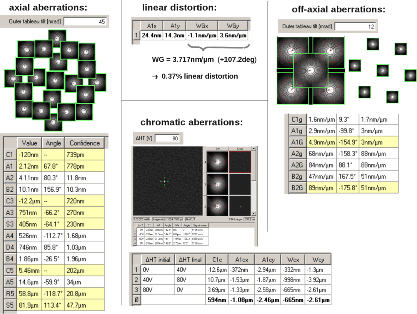
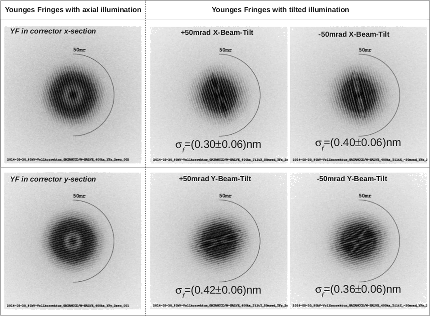
Fig. 2 shows that focus spread has a negligible effect on the resolution limit.
In February 2011, when the SALVE II phase was started, the effect of Johnson-Nyquist-noise in the electron optical elements on resolution was not well understood. The appearance of the additional image spread (for more information see here on the SALVE website) was an inexplicable phenomenon, which caused a substantial problem for the operation of CC corrected electron microscopes. It limited the atom contrast of the test material graphene for the SALVE II microscope to 2% (July 2013). After the discovery of the interrelation of Johnson-Nyquist-noise and image spread by SALVE partner CEOS GmbH Heidelberg in 2013 [3], the electron optical components of the corrector have been appropriately redesigned to improve its performance.
Reduction of image spread due to Johnson-Nyquist-noise in the CC/CS corrector
Tests at CEOS GmbH in Heidelberg on the upgraded corrector for the chromatic aberration corrected PICO-instrument of the research center Jülich (CCOR+) had already shown that the effect of Johnston-Nyquist-noise can be significantly reduce by a reduction of the beam diameter in the corrector accompanied by an increase of the magnetic and electric field strengths. The increase of the electric field strengths of the multipoles from 5 to 8 kV is necessary in order to maintain their correction power. The strength of the magnetic multipoles was increased accordingly in the SALVE corrector.
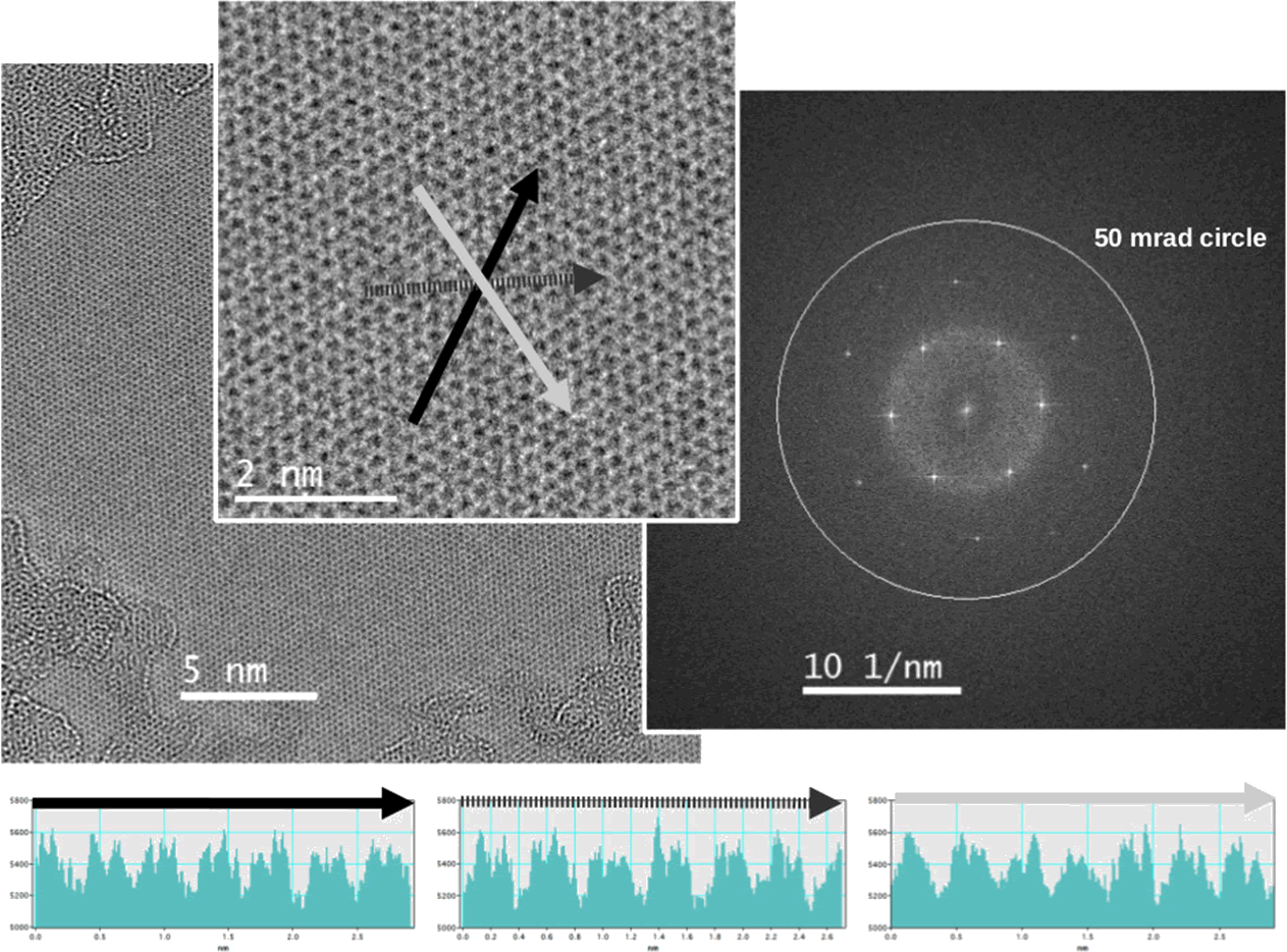
These measures have reduced the image spread caused by the corrector from 35 pm to 25 pm. At present an image spread (see research/instrument development) of 35 pm was measured at 80 kV with the SALVE II instrument (corrector 25 pm and Zeiss Libra column 25 pm). Recent results of the SALVE II+ corrector convincingly show that the electron optical design enables phase contrast imaging with a hitherto not-existing quality. As an example, Fig. 3 shows the phase contrast image of graphene taken at 80 kV. Here, the individual carbon atoms are resolved. Contrast transfer is shown up to about 8% above background and frequency transfer until 106 pm (August 2014).
Summary
A new CC/CS-corrector optimized for operation between 20 and 80 kV has been developed in two stages (SALVE II and SALVE II+) and integrated in an transmission electron microscope. During the development phase of SALVE II, a strong contrast-limiting effect in Cc/Cs correctors designed with varying magnetic and electrostatic fields has been discovered, that is the Thermal Magnetic Field Noise. It was experimentally verified and theoretically understood [3]. Although it is not possible to eliminate this effect without conducting He-cooled microscopy, in the SALVE II+ stage Johnson-Nyquist-noise is reduced and contrast transfer is reaching up to 50 mrad.
References
- Rose, H. (1992). Prospects of aberration corrected TEM. 10th Eur. Congr. El. Micr. (Granada, Spain), 47
- Rose, H. (1992). Electron optical correction system for lenses of electron microscope - has series of electric or magnetic and electromagnetic quadrupole elements located in optical path in astigmatic intermediate images. Patent, DE4204512 A1
- Uhlemann, S., Müller, H., Hartel, P., Zach, J., & Haider, M. (2013). Thermal magnetic field noise limits resolution in transmission electron microscopy. Physical review letters, 111: 046101, doi: 10.1103/PhysRevLett.111.046101
- Schmidt, Th., Groh, U., Fink, R., Umbach, E., Schaff, O., Engel, W., Richter, B., Kuhlenbeck, H., Schlögel, R., Freund, H. J., Bradshaw, A. M., Preikszas, D., Hartel, P., Spehr R., Rose, H. H., Lilienkamp, G., Bauer, E., & Benner, G. (2002) XPEEM with energy-filtering: advantages and first results from the SMART project. Surface review and letters, 9: 223-232, doi: 10.1142/S0218625X02001811